Diagram Label And Explain The Components Of A Muscle Fibres
Labeling tissue endomysium perimysium epimysium fibers connective Mitochondria again, but this time in muscle! Muscular system anatomy physiology nurseslabs muscle fibers body cells gross called myosin made human nursing some together school exercise choose
Diagram of Muscle Fiber: 3 Types, Functions, and Anatomy - WOMS
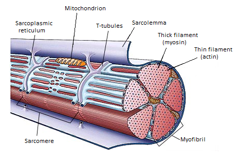
Muscle fibers types will Types of muscle tissue and fibers Skeletal fibers sarcomere myofibril labeled sarcolemma membrane physiology plasma
Muscle fibers structure biology ib
#121 striated musclesDiagram of muscle fiber: 3 types, functions, and anatomy Muscle fibers filament sarcoplasmic skeletal myofibril thick section cross structure muscles hypertrophy below following organizationSkeletal muscle.
Diagram of muscle fiber: 3 types, functions, and anatomyStructure of skeletal muscle Muscle diagram anatomy saved fiber owensboro kctcs legacy edu skeletalMuscle fiber structure and inner parts anatomical description outline.

Parts of a muscle fiber quiz
Structure of a skeletal muscle fiber diagramStructure of muscle fibers (ib biology) Muscle fibre: learn definition, properties and factsLeg muscle anatomical structure, labeled front, side and back view.
Muscle fiber anatomyLabel the structures of a muscle fiber using google slides Skeletal muscle fiber labeled diagramMuscle structure skeletal fiber cell anatomy muscular seer training single illustration.

Single muscle fiber diagram
Muscle structure skeletal gross fiber ultra whichSkeletal muscle organization Muscle cell skeletal fibers fiber figure membrane tissue cytoplasm biology called plasma sarcolemma sarcoplasm myofibrils appearance long fibrils types composedMuscle skeletal anatomy cell muscles muscular fibre contraction physiology human.
Striated muscles muscle fibres syncytium structure level biology cells specialized called made3,451 contraction musculaire royalty-free photos and stock images Muscle fibers explained: type i and type ii (slow & fast twitchSkeletal muscle structure layers with anatomical arm closeups outline.

Muscle fiber diagram unlabeled png image
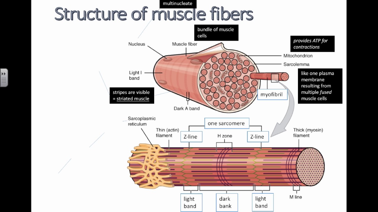
Question 6diagrammatically show the difference between the three typesSkeletal muscle Muscle cell skeletal fiber biology membrane cytoplasm sarcolemma called surrounded myofibrils figure fibers sarcoplasm illustration 2e openstax tubular plasmaMuscular system anatomy and physiology.
Cumulative topic 6: microanatomy of myofiberMuscular system anatomy physiology nurseslabs muscle fibers body cells gross called myosin made nursing some school exercise human also study Muscle skeletal contraction tissue histology teachmephysiologyMuscular system anatomy and physiology.

Muscle-skeletal muscle-gross and ultra structure
Muscle contraction and locomotionMuscle skeletal sarcolemma fibers anatomy fiber structure labeled myofibril sarcomere parts single sarcoplasm membrane figure which plasma 10.2 skeletal muscle – anatomy & physiology.
.

STRUCTURE OF A SKELETAL MUSCLE FIBER Diagram | Quizlet

Skeletal Muscle | Anatomy and Physiology I
Structure of Muscle Fibers (IB Biology) - YouTube

cumulative topic 6: microanatomy of myofiber | Skeletal muscle anatomy

Diagram of Muscle Fiber: 3 Types, Functions, and Anatomy - WOMS

Single Muscle Fiber Diagram | Quizlet

Muscle Fibers Explained: Type I and Type II (Slow & Fast Twitch